Figure 1.
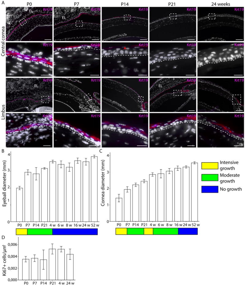
Krt19 expression pattern correlates with cornea growth and maturation. Krt19 expressing cells are marked with magenta. (A): At P0, all corneal epithelial cells were Krt19+. Boxes indicate the location of insets. At P7, the expression domain excluded the basal cells in the central and limbal cornea. The border between epithelium and stroma is marked with a dashed line. By P14, the central corneal epithelial cells were almost all Krt19-negative (arrowhead), but the limbal epithelial cells were Krt19+. At P21, we could not detect Krt19 expression in the central cornea anymore, in contrast to noticeable expression in the limbus. After that the expression pattern remained constant; at 24 weeks of age the limbal epithelial cells were Krt19+. However, signal from the central and peripheral cornea was completely lost. (B-C): Growth rate is indicated with a color pattern: yellow for intensive growth, green for moderate growth and blue for no more growth. (B): Eyeball diameter increased quickly from P0 until P7 (p=0.000), then with moderate pace from P7 until 4 weeks (p=0.000); after that growth ceased (p ≥ 0.474). (C): Cornea diameter increased stepwise between P0 and P7 (P=0.000) as well as between P21 and 4 weeks (p=0.005), with periods of moderate growth (p=0.000) after each quick growth phase. Cornea diameter did not increase anymore after 16 weeks of age (p ≥ 0.193). (D): The amount proliferating, Ki67+ cells were steadily maintained in an appropriate level at each age (p ≥ 0.125). Errors bars indicate 2 ± standard error. Hoechst for nuclear staining (white). Eyelid (EL). Scale bars are 100 µm, 20 µm for insets.

