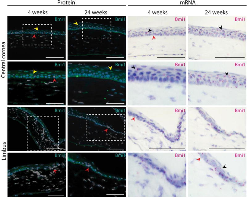
Figure 3.
Bmi1 expression pattern remains unchanged after cornea maturation. We assayed both BMI1 protein (in turquoise) and Bmi1 RNA (in magenta). Young (4 weeks old) and adult mice (24 weeks old) displayed the same expression pattern. Most of the epithelial basal cell layer of the cornea was composed of Bmi1+ cells. In both stages, we discovered few Bmi1-negative cells in the basal layer (red arrowhead). Some suprabasal cells expressed Bmi1 in the adult cornea (yellow or black arrowhead). Hoechst for nuclear staining (white) and hematoxylin for nuclear staining (blue). Scale bars are 100 µm, 50 µm for the insets.

