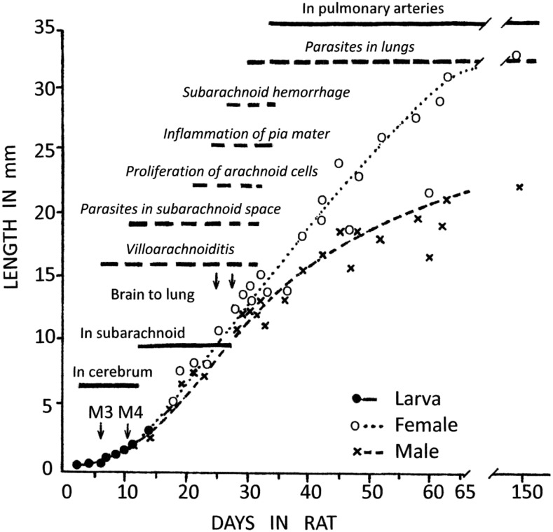
Figure 1.
Composite incorporating Figure 13 of Mackerras and Sandars4 (solid bars, normal text), depicting growth of Angiostrongylus cantonensis larvae in rat brain, with superimposed pathological findings from Figure 1 in Jindrak7 (broken bars with italicized text). M3 and M4 indicate the two larval molts in the central nervous system, beyond which the sexes are readily distinguishable; adult (L5) females grow faster and longer than males. Note the closely similar timing of both subarachnoid and pulmonary stages in both sources, given that the first study actually used A. mackerrasae.

