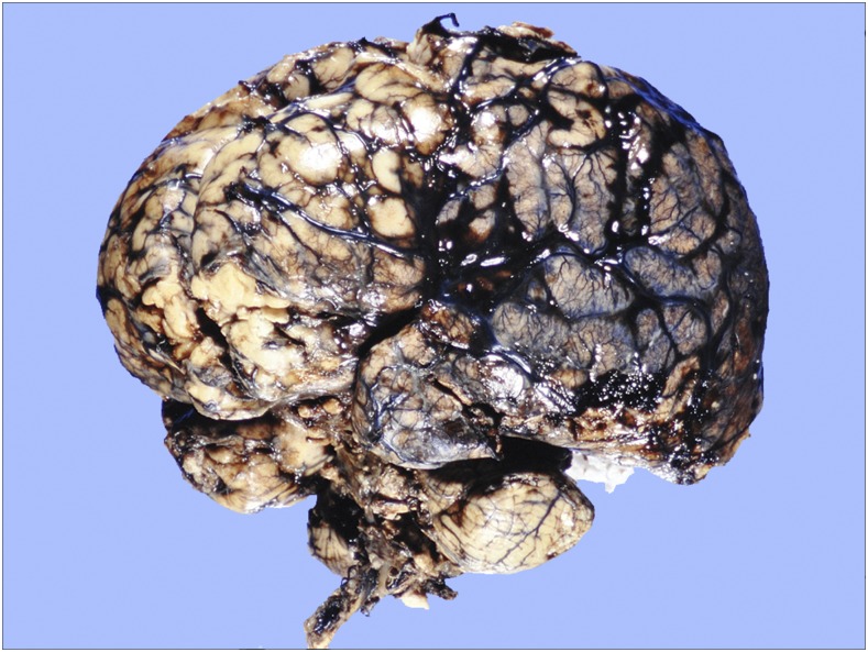
Figure 2.
Case 1: Intact brain, cerebellum, and brainstem after formalin fixation. Note vascular congestion and patchy subarachnoid hemorrhages over cerebral convexities, with generalized broadening of gyri and narrowing of sulci. Occasional worms were present on surfaces, but not evident at this magnification. This figure appears in color at www.ajtmh.org.

