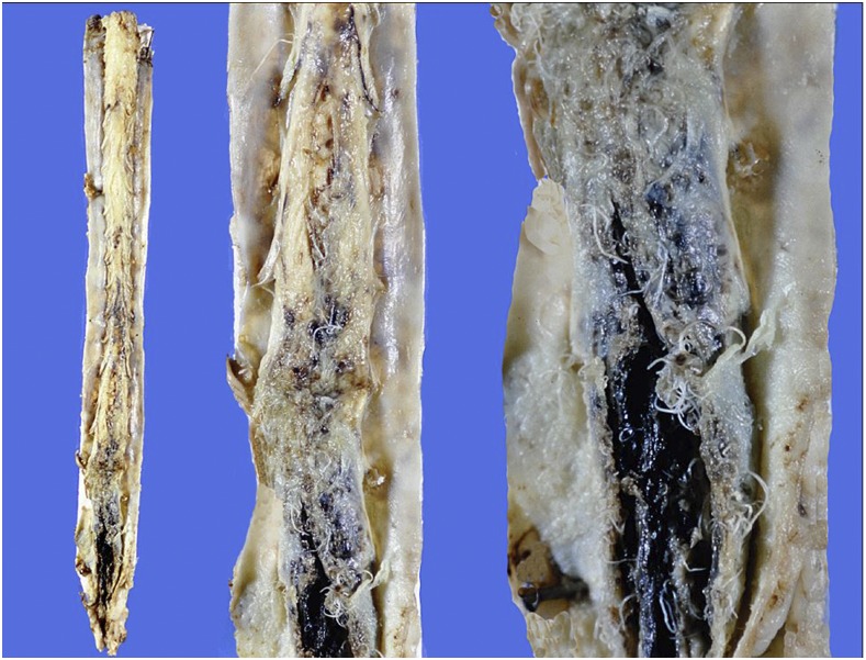
Figure 3.
Case 1: Triptych showing entire spinal cord (left) and increasing magnifications of cauda equina region (center and right). Note dense subarachnoid hemorrhage, with worms enmeshed among nerve roots and protruding into subdural space. This figure appears in color at www.ajtmh.org.

