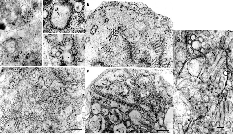
Figure 1.
Ultrastructure of Kampung Karu (KPKV), Long Pine Key (LPKV) and La Tina (LTNV) viruses in C6/36 cells. (A) KPKV. Accumulations of virions ∼40 nm in diameter inside cytoplasmic vacuoles (arrows). Bar = 100 nm. (B) KPKV. Two virions inside a cistern of granular endoplasmic reticulum (arrow). Bar = 100 nm. (C) KPKV. Smooth membrane structures (SMS) inside an expanded cistern of granular endoplasmic reticulum (arrowheads) or in a tightly apposed cistern (double arrowhead). Bar = 100 nm. (D) LPKV. Virions (arrowheads) and SMS inside an enormous expansion of a cistern of granular endoplasmic reticulum. Thick arrow indicates cross sections of the SMSs. Virions can also be found inside individual small vacuoles (thin arrows). N-fragment of host cell nucleus. Bar = 0.5 µm. (E) LPKV. Virions (thin arrows) and SMS inside an enormous expansion of granular endoplasmic reticulum. Thick arrow indicates cross sections of SMSs. Bar = 0.5 µm. (F) LPKV. Virions (thin arrows) and SMS inside an enormous expansion of granular endoplasmic reticulum. Virions can be also observed inside individual vacuoles (arrowheads). Some SMS can be very long, up to 2.2 µm (thick arrow). N-fragment of the host cell nucleus. Bar = 0.5 µm. (G) LTNV. Virions (arrowheads) and SMS (arrows) inside an expanded cistern of granular endoplasmic reticulum. Bar = 100 nm.

