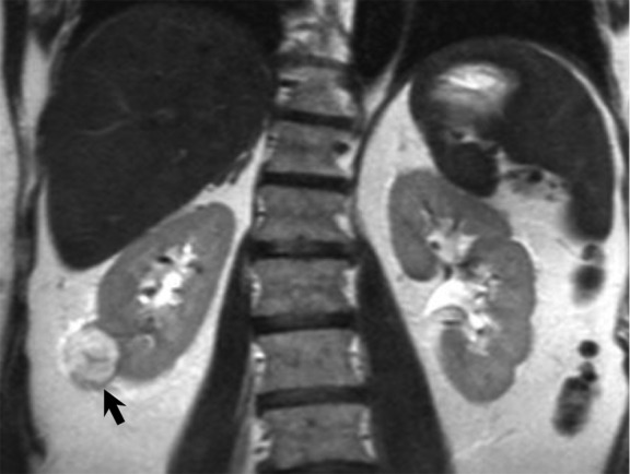
Figure 3a:

MR images show clear cell renal cell carcinoma (RCC). (a) Coronal non–fat-saturated T2-weighted single-shot fast spin-echo MR image shows a high-signal-intensity exophytic small renal mass (arrow) in the lower pole of the right kidney. (b) Coronal contrast-enhanced (corticomedullary) fat-saturated T1-weighted gradient-recalled-echo MR image shows the lesion (arrow) has high contrast enhancement. Axial (c) in-phase and (d) out-of-phase non–fat-saturated T1-weighted gradient-recalled-echo MR images. Note the presence of intravoxel fat, characterized by signal dropout, on d in comparison with c (arrows). Surgical resection enabled confirmation of clear cell RCC.
