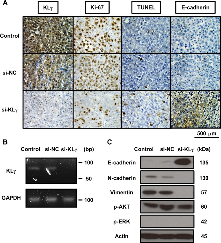
Figure 4.
(A) Representative images of resected xenografts from each treatment group stained with four immunological markers. The expression levels of KLγ, Ki-67, TUNEL, and E-cadherin are noted. Expression levels of each marker in tumors of mice treated with KLγ siRNA compared to both the control group and the group of mice treated with negative control siRNA. The expression levels of KLγ and Ki-67 decreased in tumors of mice treated with KLγ siRNA. The expression levels of TUNEL and E-cadherin increased in tumors of mice treated with KLγ siRNA. (B) Treatment with KLγ siRNA suppressed the expression levels of KLγ mRNA in tumors of mice treated with KLγ siRNA, as measured by RT-PCR analysis. (C) Western blot analysis of protein extracted from resected xenografts in each treatment group. The expression levels of E-cadherin, N-cadherin, vimentin, phospho-AKT, phospho-ERK1/2, and actin (as a control) were noted. The expression level of E-cadherin was increased in tumors of mice treated with KLγ siRNA. The expression levels of N-cadherin and vimentin decreased in tumors of mice treated with KLγ siRNA.

