
Figure 1. Statins enhance the sensitivity to MEK inhibitors.
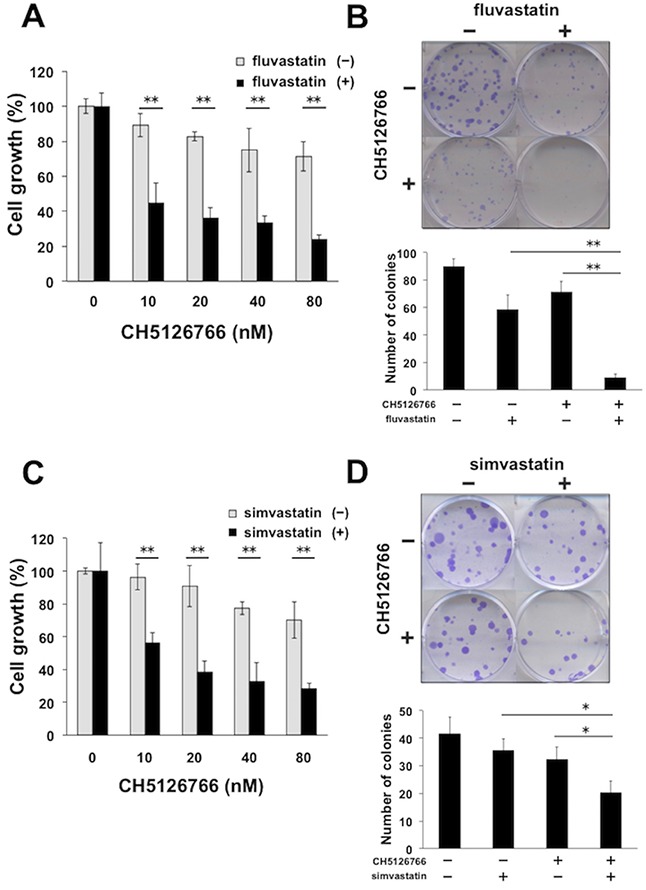
(A) Growth inhibitory effects of CH5126766 with or without fluvastatin on MDA-MB-231 cells. Cells were treated with CH5126766 at the indicated concentrations with or without fluvastatin (0.3 μM) for 72 h, and the cell viability was measured with a Cell Counting Kit-8 assay. Data obtained with DMSO with or without fluvastatin was taken as 100%. Columns, means of triplicate data; bars, standard deviation (SD); **, P < 0.01. (B) Suppression of colony formation by the combined treatment of CH5126766 with fluvastatin. MDA-MB-231 cells were treated with CH5126766 (40 nM) and/or fluvastatin (0.3 μM) for 72 h. After further incubation for colony formation with fresh medium, the number of colonies was counted. (upper panel) The representative images of stained colonies are shown. (lower panel) Colony numbers are shown in the graph. Columns, means of triplicate data; bars, SD; **, P < 0.01. (C) Growth inhibitory effects of CH5126766 with or without simvastatin on MDA-MB-231 cells. Cells were treated with CH5126766 at the indicated concentrations with or without simvastatin (0.3 μM) for 72 h, and the cell viability was measured with a Cell Counting Kit-8 assay. Data obtained with DMSO with or without simvastatin was taken as 100%. Columns, means of triplicate data; bars, standard deviation (SD); **, P < 0.01. (D) Suppression of colony formation by the combined treatment of CH5126766 with simvastatin. MDA-MB-231 cells were treated with CH5126766 (20 nM) and/or simvastatin (0.3 μM) for 72 h. After further incubation for colony formation with fresh medium, the number of colonies was counted. (upper panel) The representative images of stained colonies are shown. (lower panel) Colony numbers are shown in the graph. Columns, means of triplicate data; bars, SD; *, P < 0.05.