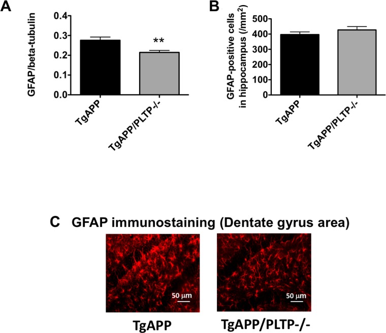
Figure 5. Quantification of astrogliosis in TgAPP and TgAPP/PLTP–/– brain tissues.
(A) The expression of the astrocyte activation marker GFAP was quantified in brain homogenates from 6 months old TgAPP (n = 7) and TgAPP/PLTP–/– mice (n = 6) by Western Blot. (B) Tissue sections from TgAPP (n = 5) and TgAPP/PLTP–/– mice (n = 5) were stained with anti-GFAP antibodies to detect astrogliosis and observed by fluorescence microscopy. For each animal, GFAP-positive cells were counted in 3 fields (1 in the dentate gyrus, 1 in CA1, 1 in CA3) from 4 different sections/animal. **p < 0.02 vs TgAPP mice (Student’s t test). (C) Representative images of the staining observed in each group.

