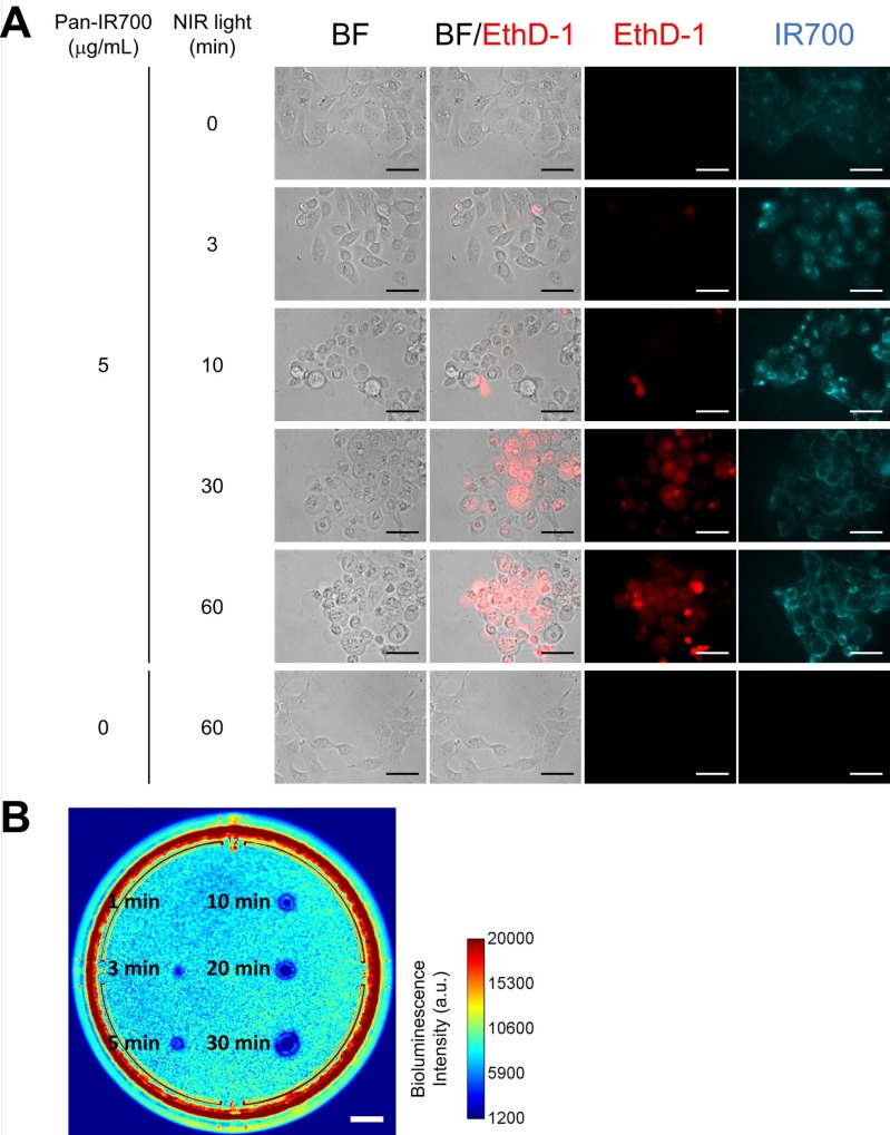
Figure 2. In vitro NIR-PIT for A431-GFP-luc cells.
(A) Phase and fluorescent microscopy of NIR-PIT treated A431-GFP-luc cells, which were pre-incubated with Pan-IR700 (5 μg/mL) at 37° C for 1 h. NIR-PIT induced cell death with cell swelling and bleb formation. EthD-1 staining showed cell death in a light dose-dependent manner. Scale bar: 50 μm (original magnification, 40×). (B) Bioluminescence imaging of a 10 cm dish demonstrated that luciferase activity in A431-GFP-luc cells decreased with increasing light dose. Scale bar: 1 cm.

