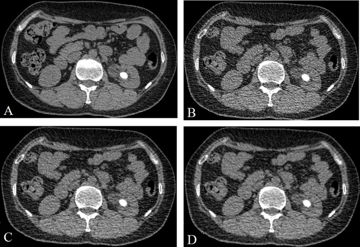
Figure 1. A 48 years old man with a stone (14.2 mm × 10.3 mm × 13.1 mm) in the left kindy.
(A) CDCT images, reconstructed with FBP; (B) LDCT image, reconstructed with FBP; (C) LDCT image, reconstructed with 60% ASIR; (D) LDCT images, reconstructed with 80% ASIR. The stone size were no obvious difference between the A and D.

