
Figure 1.
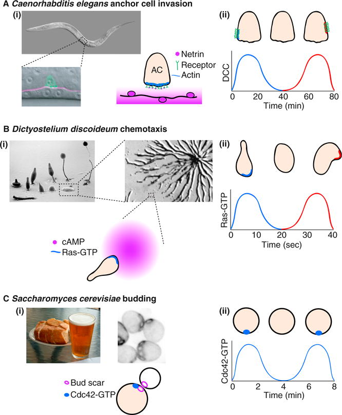
Removal of spatial cues reveals a potential for oscillatory behavior in several polarity systems.
(Ai) The C. elegans anchor cell (AC) invades through the underlying basement membrane to initiate uterine–vulval attachment (pictures courtesy of Judith Kimble, University of Wisconsin-Madison, and [17]). The anchor cell polarizes towards the spatial cue, netrin, which is released by the ventral nerve cord. (Aii) In animals without netrin, the anchor cell netrin receptors cluster at a random location, disperse, and then repolarize at a different site. The graph represents the concentration of netrin receptor (UNC-40/DCC) at the blue and red sites in the picture [1]. (Bi) D. discoideum cells migrate up the cAMP gradient released by other cells as they aggregate (pictures courtesy of M.J. Grimson and R.L. Blanton, Texas Tech University, and [18]). (Bii) In uniform cAMP, the cells polarize transiently, then depolarize and repolarize in a random direction. The graph represents the local concentration of the polarity regulator Ras-GTP [7]. Note the faster time scale of clustering and disassembly. (Ci) S. cerevisiae cells polarize towards an internal spatial cue at the bud scar. (Cii) In mutants lacking the spatial cue, cells cluster Cdc42 at a random location in an oscillatory manner. The graph represents the local concentration of Cdc42-GTP in the cluster [8].