Fig. 3.
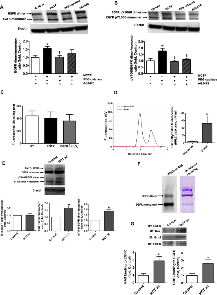
Covalent dimerization of EGFR increases EGFR signaling in vitro and in vivo. The levels of EGFR dimer and monomer in PASMC co-cultured with MCTP pretreated PAEC were obtained by performing Western blot analysis of PASMC lysates. The presence of SDS-resistant an EGFR dimer was significantly increased in PASMC co-cultured with PAEC pretreated with MCTP and this was attenuated by pretreatment with PEG-catalase (250 U/ml), but not by the EGFR inhibitor AG1478 (100 nM, A). The ratio of auto-phosphorylated EGFR dimer/auto-phosphorylated EGFR monomer was also significantly increased in PASMC co-cultured with PAEC pretreated with MCTP and attenuated by PEG-catalase or the EGFR kinase inhibitor, AG1478 (B). HEK cells over-expressing EGFR were treated with CuCl2/H2O2 (10 µM/300 µM) to induce oxidative stress. Whole cell lysates were then examined for specific dityrosine fluorescence. Global tyrosine oxidation was unchanged (C) but HPLC analysis of the peptide fragments from dimer and monomer bands of immunoprecipitated and digested EGFR revealed the presence of dityrosine crosslinks only in the EGFR dimer (D). Western blot analysis revealed no change in total EGFR protein levels in peripheral lung tissue lysates prepared from control and MCT-treated rats at 3 days (E, left panel). Loading was normalized to β-actin. However, the EGFR dimer/monomer ratio (E, middle panel) and the levels of auto-phosphorylation, assessed by measuring pY1068EGFR (E, right panel), were both significantly in MCT treated rats. The levels of EGFR dimer assessed by Western blot analysis of EGFR immunoprecipitation (F, left panel) appear to be much less when compared to the band present with Coomassie staining (F, right panel). The paradox may be due to reduced ability of antibodies to recognize the covalently cross-linked EGFR protein. Thus, the amount of EGFR dimer and its contribution into uncontrolled cell growth may be underestimated based on the data obtained by Western blot. The level of EGFR signaling was evaluated by immunopreciptating EGFR and probing for Grb2 and Ras. EGFR/Grb2 and EGFR/Ras interactions were significantly increased in MCT-treated rats (G). Results are expressed as mean±SEM; N=3–7. *P<0.05 vs. control group; †P<0.05 vs. PASMC co-cultured with MCTP treated PAEC.

