
Figure 1.
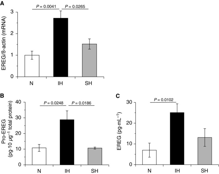
IH increased the expression of EREG in hCASMCs. hCASMCs were cultured for 24 h with serum‐free medium and exposed to normoxia, IH, or SH for 24 h. (A) Total RNA were extracted, and real‐time RT‐PCR was performed using specific primers for human EREG mRNA, as described in the Materials and methods section. Each value was normalized by arbitrarily setting the value of β‐actin of the cells exposed to normoxia to 1.0. The results are representative of four independent experiments. (B) Normoxic‐, IH‐, and SH‐treated hCASMCs were denatured by repeats of freeze and thaw. Ten micrograms of total protein in each cell lysate was used in an EREG immunoassay, as described in the Materials and methods section. The results are representative of five independent experiments. (C) Conditioned media of normoxic‐, IH‐, and SH‐treated hCASMCs were collected and used in an EREG immunoassay, as described in the Materials and methods section. The results are representative of four to five independent experiments. Each point represents the mean ± SEM.