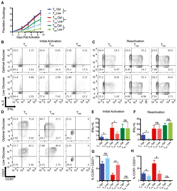
Figure 2. Effector Memory T Cells Are Resistant to Glucose-Mediated IFN-γ Suppression.
(A) The indicated subsets were stimulated with anti-CD3/CD28 coated beads in the presence of optimal (35 mM) or low (0.35 mM) glucose. Cell expansion was monitored by Coulter counter on the indicated days. Statistics were performed on day 9 population doublings.
(B and C) T cell subsets that were expanded for 24 hr (B) with anti-CD3/CD28-coated beads or expanded for 9–11 days (C) with anti-CD3/CD28-coated beads before IFN-γ/IL-2 production was measured after PMA/ionomycin treatment.
(D) CCR7 and CD27 expression measured on T cell subsets described in (A) 7 days after T cell expansion.
(E and F) IFN-γ production by cells from the initial activation (E) and reactivation (F) are summarized from three independent experiments and donors (see Figure S3 for IL-2 and TNF-α quantification).
(G and H) Quantification of CCR7+ CD27+ (G) and CCR7– CD27– (H) cells are summarized from three independent experiments.
Error bars reflect SEM. *p < 0.05, **p < 0.01, paired two-tailed Student’s t test. ns, not significant.

