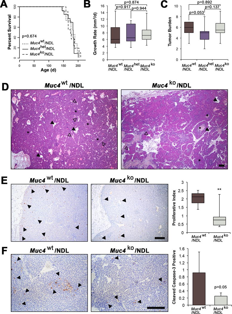
Figure 2. Muc4 deletion modestly alters primary mammary tumor histology but does not affect mammary tumor latency or growth rate in the NDL model.

(A-C) Survival curves and box plots depicting Muc4/NDL tumor growth characteristics. Latency to tumor formation (A), tumor growth rate (B), and the overall tumor burden (number of glands with tumors) (C), were not significantly different among genotypes (Muc4wt/NDL [n=13], Muc4het/NDL [n=14] and Muc4ko/NDL [n=12]). Data are presented as means ± SD and significance determined by log-rank test (A) or 2-sided Students-t test (B-C). (D) Representative H&E images are depicted of formalin fixed, paraffin embedded sections from Muc4wt/NDL and Muc4ko/NDL primary tumors. Tumors from both genotypes develop as heterogeneous mammary adenocarcinomas displaying nests of tumor epithelium (asterisks) and abundant lumina (closed arrowheads) that are structurally distinct from endothelial lined blood vessels (open arrowheads). (E) Representative images Muc4wt/NDL and Muc4ko/NDL primary tumor tissues following immunodetection of the proliferation marker phospho-histone H3. Quantification of at least six biological replicates revealed a significant reduction in the number of proliferating cells with loss of Muc4 (**, P<0.01; t-test). The percent of pH3 positive cells/total cells is plotted. Examples of positive nuclei are denoted by closed arrowheads. (F) Immunodetection of cleaved caspase-3 suggests a modest yet insignificant reduction in the rate of apoptosis with Muc4 loss (P=0.05; t-test). Data are averages from at least five biological replicates and expressed as the percent of total cells positive for cleaved caspase-3 ± SD, where examples of positive cells are indicated with closed arrowheads. Scale bars in all images = 250μm.
