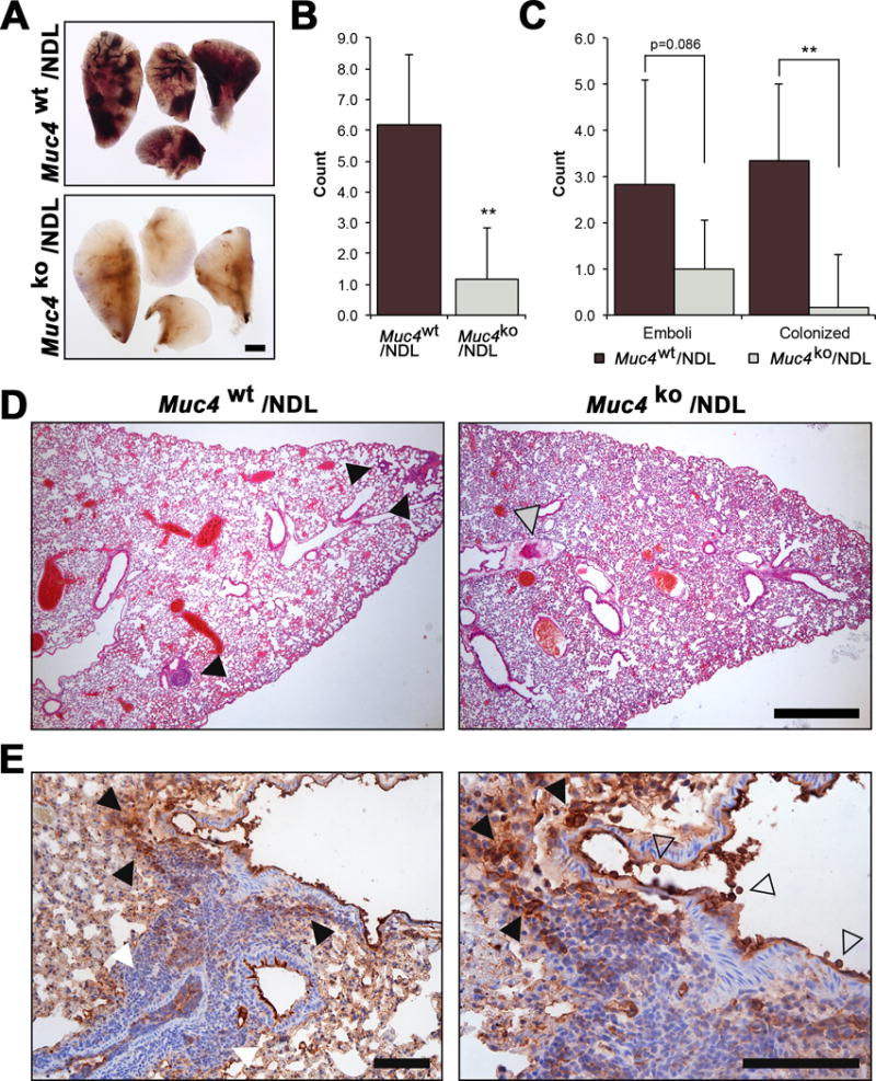
Figure 5. Muc4 deletion impairs the metastatic capacity of tail vein injected NDL cells.

Pooled (n=4 independent biological sources per line) primary Muc4wt/NDL and Muc4ko/NDL tumor cells were instilled into the lateral tail vein of FVB wt mice (n=6). Tissues (lungs, digestive tract, liver, spleen, uterus, brain) were collected 28d later, fixed in 4% paraformaldehyde and evaluated for the appearance of gross metastasis. Only lungs were found to contain gross lesions, more readily visible by carmine alum stain (A). Scale bar = 1mm. (B-D). H&E stained sections were used to quantify the occurrence of metastatic lesions. The total number of metastases (B) and the frequency of colonized lesions (C) formed in the lungs of mice injected with Muc4wt/NDL cells was significantly increased compared to those receiving Muc4ko/NDL cells. There was a modest reduction in the frequency of emboli formed by Muc4ko/NDL cells (P = 0.086). Representative H&E stained sections are presented in (D). Closed grey arrowhead, emboli; closed black arrowhead, colonized lesion. Scale bar = 1mm. (E) The level of Muc4 expression within a colonized lesion is heterogeneous, where it is strong both within (open arrowhead) and around the vascular lumen (closed black arrowhead) but is decreased in tumor cells found further away from this region (closed white arrowhead). Tissue was stained with an antibody to Muc4 beta, as described in Figures 1B and 1C. Scale bar = 150μm.
