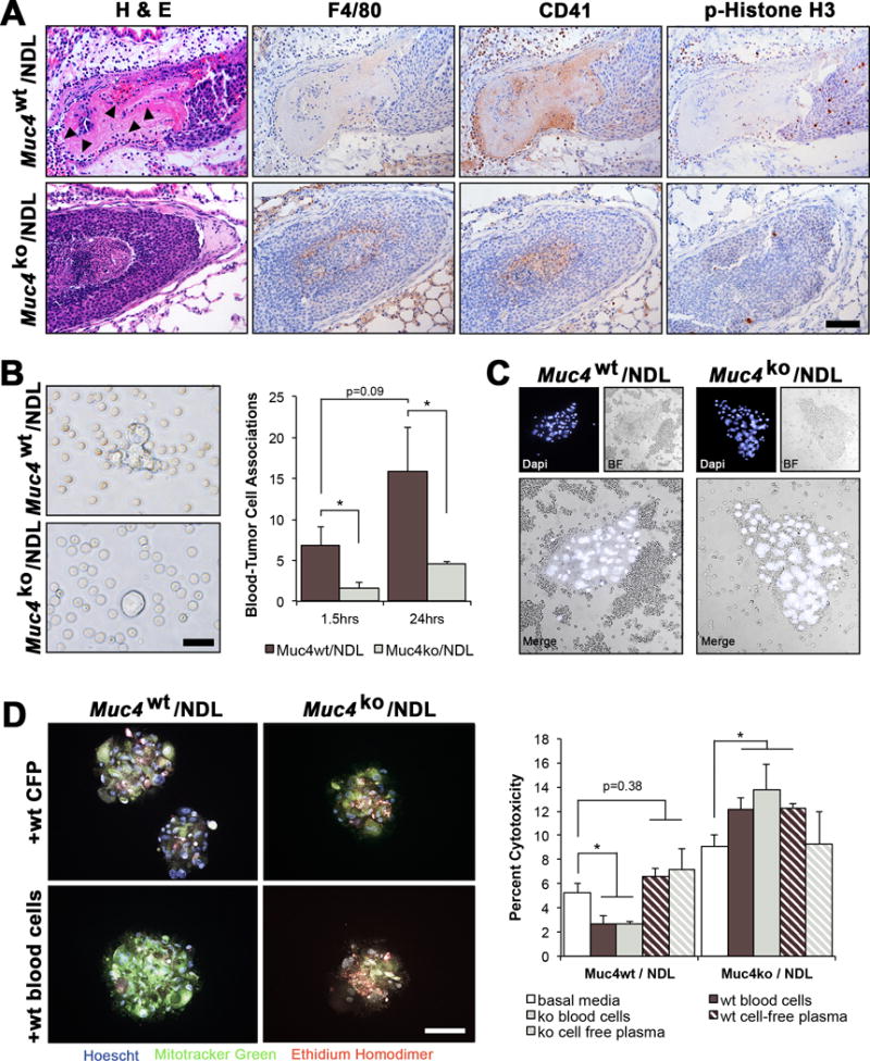
Figure 6. Deletion of Muc4 suppresses the association of tumor cells with red and white blood cells in vitro and in vivo.

(A) Representative H&E stained lung lesions from Muc4wt/NDL and Muc4ko/NDL mice, illustrating the elevated abundance of blood cells (closed arrowheads) in Muc4wt/NDL emboli. Immunodetection of proteins specific to macrophages (F4/80) and platelets, megakaryocytes and hematopoietic progenitors (CD41/Integrin alpha 2b) indicated the majority of associated blood cells to be CD41 positive. Phospho-histone H3 (pH3) staining revealed Muc4wt/NDL lung lesions were more proliferative than Muc4ko/NDL lesions. Scale bar in all images = 100μm. Images are representative of at least 6 biological replicates. (B) Quantification of tumor cell-blood cell associations (proximity of < 5μm) using bright field microscopy of live cells revealed a reduced number of associated blood cells at either 1.5hrs or 24hrs after the initiation of co-culture conditions. Pooled NDL cells (n=4 biological sources per line) were used for these experiments and data are presented as averages of three independent experiments ± SD. Scale bar = 50μm. *, P < 0.05. (C) Similar associations were noted for Muc4wt/NDL cells in vivo, where the abundance of closely associated blood cells appears visibly enhanced by the presence of Muc4. Pooled Muc4wt/NDL or Muc4ko/NDL cells (n=4 biological sources per line) were instilled into the lateral tail vein of female mice (n=4 per line) and whole blood was collected by cardiac puncture 3d later and immediately fixed in 4% paraformaldehyde. Nuclei were stained with DAPI and individual cell clusters imaged by fluorescence and bright field microscopy. Scale bar = 200μm. (D) Muc4-mediated tumor cell association with blood cells promotes tumor cell viability in suspension. Primary Muc4wt/NDL and Muc4ko/NDL tumor cells (n=3 per line) were labeled with MitoFluor green, and cultured in suspension for 24hrs after admixing with basal media, freshly harvested blood cells in basal media, or cell free-plasma (CFP) in basal media harvested from Muc4wt or Muc4ko mice. Hoechst 33342 was used as a nuclear stain to discern platelets from immune and tumor cells, and MitoFluor green was used to distinguish tumor cells from immune cells. Percent cytotoxicity (mean ± SD) was determined within tumor sphere aggregates by positive staining for ethidium homodimer. Student’s t-test revealed blood cells significantly suppress the death of Muc4wt/NDL but not Muc4ko/NDL cells. The origin of the blood cells (Muc4wt or Muc4ko) did not affect outcomes. Images are presented as final merged composites. Unmerged images may be found in Supplemental Figure 4B. Scale bar = 200μm. *, P < 0.05; ***, P < 0.001.
