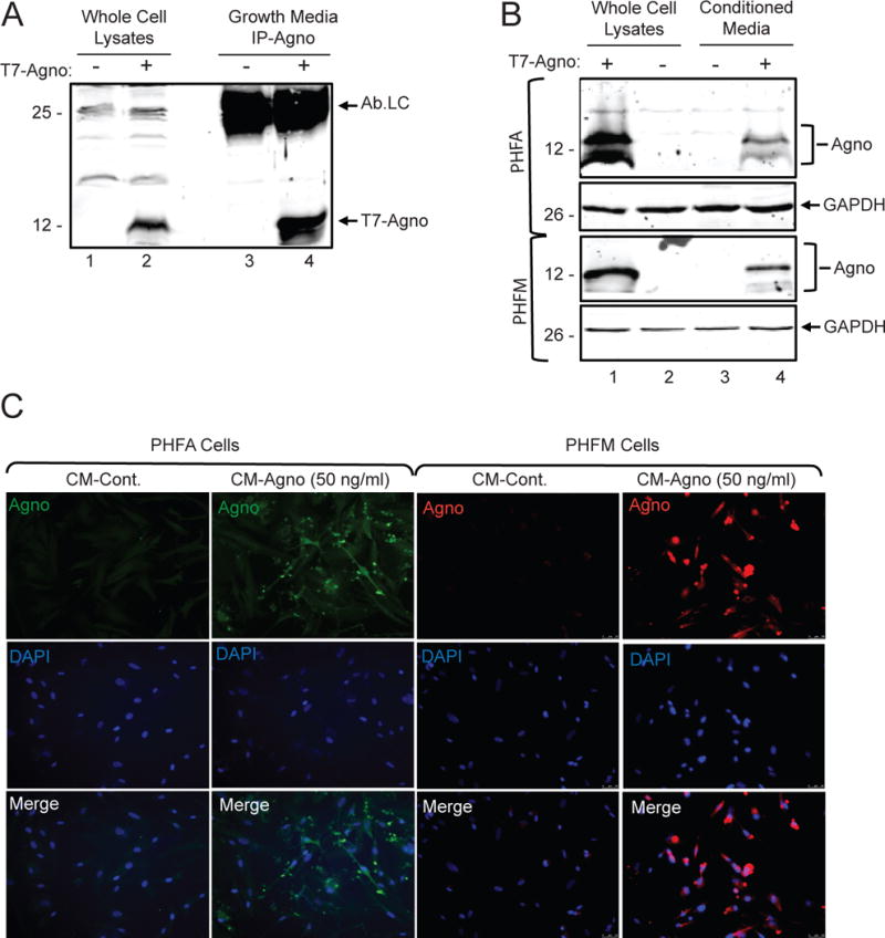
Figure 1.

Agnoprotein is released and taken up by glial cells. A. TC620 cells were transfected with pcgT7-agno and whole cell extracts and growth media was collected at 48 hours. Growth media was subjected to immunoprecipitation for the detection of agnoprotein. In lane 1 and 2, whole cell protein extracts from untransfected and transfected cells were loaded as negative and positive controls of agnoprotein expression, respectively. Ab-Lc points the light chain of agnoprotein antibody. B. Whole cell extracts of primary glial cells show uptake of extracellular agnoprotein following conditioned media treatment. Primary human fetal astrocytes (PHFA) or primary human fetal microglia (PHFM) were either transfected with pcgT7-agno or treated with CM-control or CM-agno obtained from Tc620 cells as shown in panel A. At 48 hours post-transfection and treatments, cell lysates were prepared and analyzed via Western blotting for agnoprotein and GAPDH detection. C. Immunocytochemistry analysis of primary glial cells shows uptake of extracellular agnoprotein. PHFA and PHFM cells were treated with CM-control or CM-agno. At 48 hours, cells were fixed and processed for immunocytochemistry for the visualization of agnoprotein.
