Figure 2.

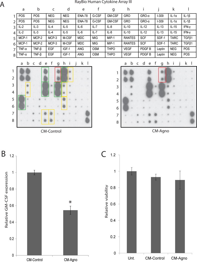
GM-CSF release by astrocytes exposed to CM-agno is suppressed. A. RayBioTech Human Cytokine Array C3 analysis of conditioned media from PHFA cells. PHFA cells were treated with CM-control or CM-agno obtained from Tc620 cells transfected with either control plasmid (CM-control) or pcGT7-agno plasmid (CM-agno) for 24 hours, after which media was changed and cells were exposed to fresh media. At 24 hours, media was collected and cytokine expression was determined using the cytokine array kit. Cytokine intensity was normalized to positive control signal intensities. Green boxes signify highly secreted cytokines, yellow boxes signify moderately secreted cytokines, and red box signify GM-CSF with a significant change in secretion following agnoprotein treatment by PHFA cells. B. Quantification of relative GM-CSF expression in the media following CM-control or CM-agno treatment. Intensities were normalized to positive control intensities from three tested membranes. C. MTT assay for the assessment of PHFA viability following treatment with CM-control or CM-agno for 24 hours. Viabilities were normalized to untreated PHFA cellular viability.
