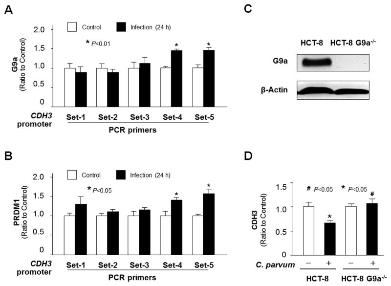
Fig. 5.
Enrichment of H3K9me3 within the CDH3 gene locus in HCT-8 cells following Cryptosporidium parvum infection involves the recruitment of G9a and PRDM1. (A–B) Increased recruitment of G9a and PRDM1 to the CDH3 gene locus in HCT-8 cells following C. parvum infection. Cells were exposed to C. parvum infection for 24 h, followed by Chromatin immunoprecipitation (ChIP) analysis using anti-G9a and anti-PRDM1, respectively, and the PCR primer sets as designed. Increased recruitment of Ga9 (A) and PRDM1 (B) was detected in the CDH3 gene locus in cells following infection. (C) Knockdown of G9a in HCT-8 cells. Cells were transfected with the G9a-CRISPR/Cas9 KO(h) and G9a-HDR plasmids; stably transfected cells were cloned and confirmed by western blot analysis. (D) Knockdown of G9a attenuated the downregulation of the CDH3 gene in cells following C. parvum infection. The CDH3 RNA levels were quantified using real-time quantitative PCR in HCT-8 and HCT-8-G9a−/− cells after exposure to C. parvum infection for 24 h. Data represent means ± S.E.M. from three independent experiments. *P<0.05, ANOVA compared with non-infected controls or empty vector controls; #P<0.05, ANOVA compared with infected controls.

