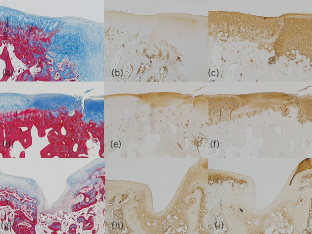
Figure 4.

Photomicrograph of the section of regenerated cartilage of the microfracture with matrix plus bone marrow aspirate concentrate group with a) Masson trichrome staining; b) immunohistochemical staining with anti-collagen type I antibody; and c) immunohistochemical staining with anti-collagen type II antibody and the microfracture with acellular matrix group (d–f); and microfracture group (x 40) (g–i), respectively
