
Figure 1.
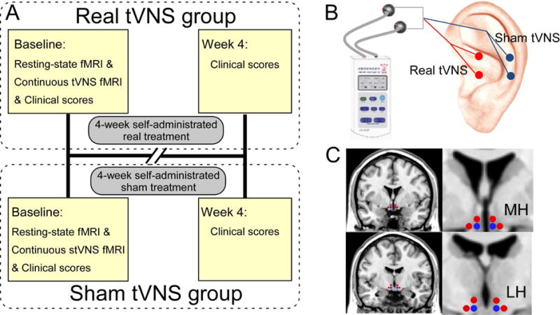
The study protocols. (A) Procedures and data used for the study. In baseline, a resting-state fMRI, a continuous tVNS fMRI and clinical scores were collected and used for the patient in the real tVNS group. All subsequent 4-week treatments were self-administered by the patients at home, except for the 6-mintue tVNS during the MRI scan. The investigators collected the patient’s clinical scores at the end of the 4-week treatment. All procedures performed in the sham tVNS treatment group were identical to the procedures for the real tVNS group, except for the location of stimulation. (B) Locations for real tVNS and sham tVNS stimulation. (C) Selected medial hypothalamus (MH) and lateral hypothalamus (LH) for seed-base whole-brain connectivity analyses (seeds marked in blue). Red dots represent the positions of controlled null seeds.