
Figure 1.
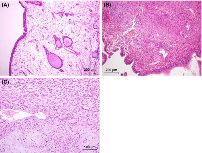
(A) Initial nasal mucosal biopsy (collected on 04 June 2013) displaying markedly edematous stroma containing low numbers of inflammatory cells and cystic mucosal glands consistent with a histopathological diagnosis of nasal polypoid hyperplasia. HE stain. (B) Interim nasal mucosal biopsy (collected on 16 October 2013) displaying area of atypical spindloid mesenchymal cells within the biopsy tissue. HE stain. (C) Third nasal biopsy (collected on 13 August 2015) displaying chondrosarcoma (bottom of field) merging with the surrounding atypical mesenchymal population (top of field). HE stain.