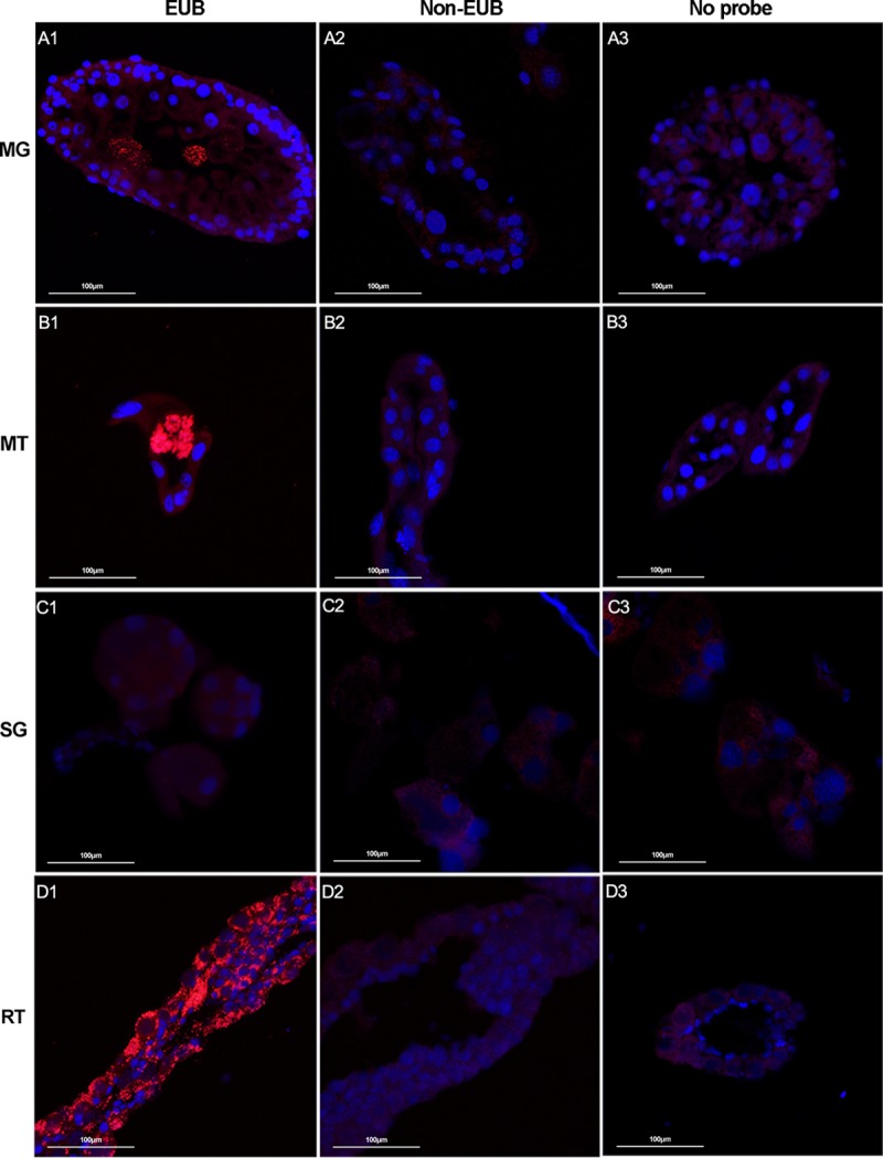
FIG 2.

Localization of microbiota in different tissues of female R. haemaphysaloides by FISH analysis with universal 16S rRNA probe (red). Nuclei were stained with DAPI (blue). Fluorescent signals were examined in midgut (A), Malpighian tubules (B), salivary glands (C), and ovaries (D). Hybridizations with noneubacterial probe (Non-EUB) and without probe were used as negative controls. Tissues from at least 5 individual ticks were pooled and used for FISH analysis. Images are representative of three independent experiments.
