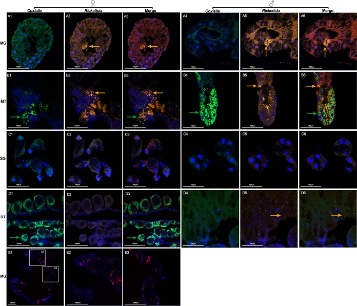
FIG 7.
Costaining of Coxiella sp. (green), Rickettsia sp. (yellow), and Pseudomonas sp. (red) in D. silvarum. Nuclei were stained with DAPI (blue). Sections of midgut (A1 to A6), Malpighian tubules (B1 to B6), salivary glands (C1 to C6), ovaries (D1 to D3), and testes (D4 to D6) were hybridized with Coxiella sp.-specific 23S rRNA probe (A1 and A4, B1 and B4, C1 and C4, and D1 and D4) and Rickettsia sp.-specific 16S rRNA probe (A2 and A5, B2 and B5, C2 and C5, and D2 and D5) simultaneously. (A3 and A6, B3 and B6, C3 and C6, and D3 and D6) Merged images of DAPI, Coxiella sp., and Rickettsia sp. staining. Green arrows denote Coxiella sp. and yellow arrows denote Rickettsia sp. MG, midgut; MT, Malpighian tubules; SG, salivary glands; RT, reproductive tissue. (E1 to E3) Whole-mount in situ hybridization of D. silvarum midgut using Pseudomonas sp.-specific 16S rRNA probe. (E2, E3) Close-up view of boxed regions in panel E1. Tissues from at least 5 individual ticks were pooled and used for FISH analysis. Each image shows a single focal plane. Images are representative of three independent experiments.

