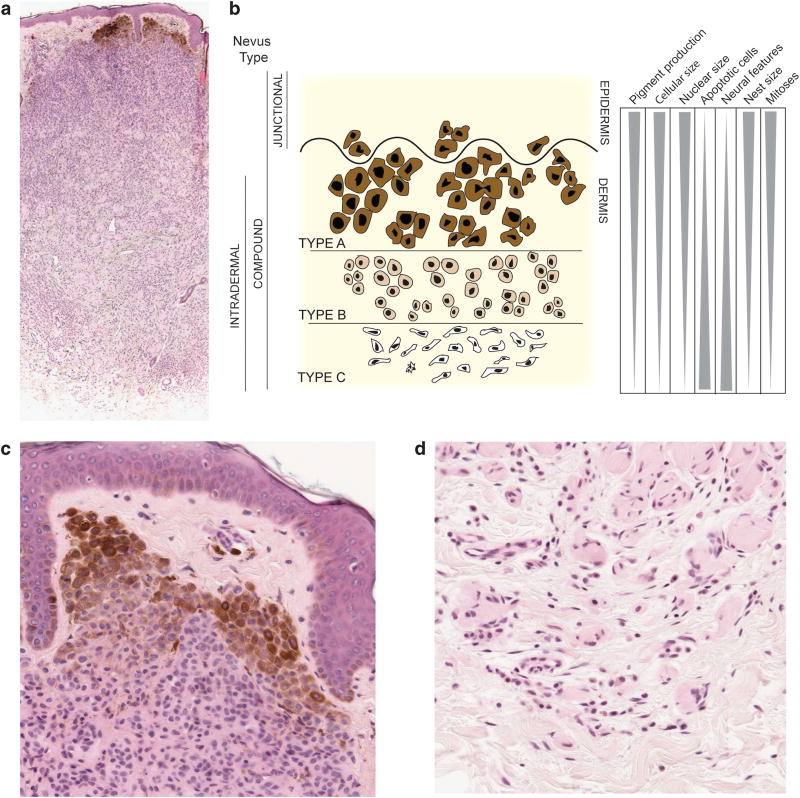
Figure 1.
Schematic of melanocytic nevus architecture. (a) Low power image of an intradermal melanocytic nevus stained with hematoxylin and eosin (H&E). The nevus shows features of maturation. (b) Junctional nevi are confined to the epidermis and appear as pigmented macules. Compound nevi have both an intra-epidermal and dermal component. Intradermal nevi are entirely confined to the dermis. Type A, B and C melanocytes are morphologically distinct and found at different depths within the skin. With increasing depth, nevi are less pigmented, smaller, have smaller nuclei, fewer mitoses, increased number of apoptotic cells and increased neural features. Nest size decreases with maturation. (c) High power images of type A melanocytes in the most superficial portion of the nevus. H&E stained section. (d) Type C melanocytes in the deepest portion of the nevus showing neural (Schwannian) differentiation. H&E stained section.

