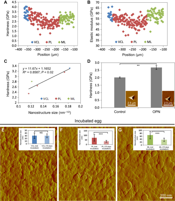
Fig. 6. Mechanical testing by nanoindentation of eggshell and synthetic calcite crystals and effects of physiologic eggshell dissolution.

(A) Hardness distribution across the eggshell layers. (B) Elastic modulus distribution across the eggshell layers. (C) Hall-Petch plot of average hardness versus nanostructure size distribution in the eggshell layers. (D) Hardness values from synthetic calcite crystals grown in the absence (control) and presence of OPN (5.9 μM). Insets show typical images of residual indents on the specimen surface. Significant difference is indicated by a bracket (**P < 0.01). (E to G) AFM images of nanostructured VCL (E), middle PL (F), and ML (G) eggshell layers from a fertilized egg incubated for 15 days (scanning area, 1.2 μm × 1.2 μm). Insets show nanostructure size distribution of the different eggshell layers, comparing eggs that were not incubated to incubated eggs. No significant difference (P > 0.05) is observed between VCLs, whereas a significant difference (***P < 0.001) in size exists between the two groups for the PL and the ML. Values were compared by a two-paired Student’s t test.
