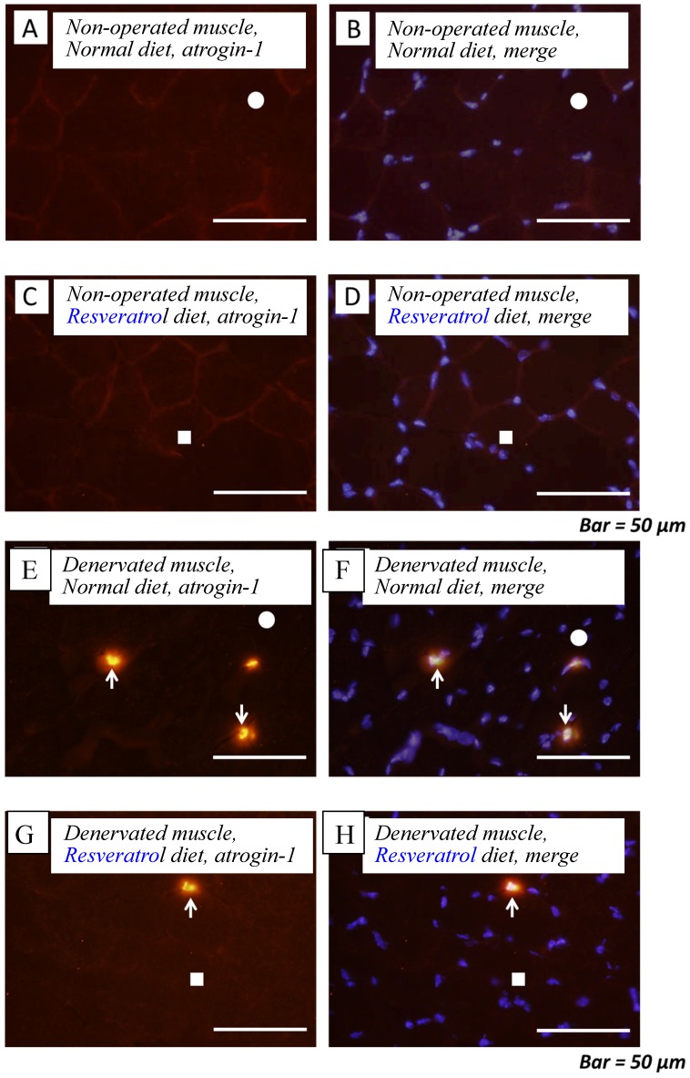
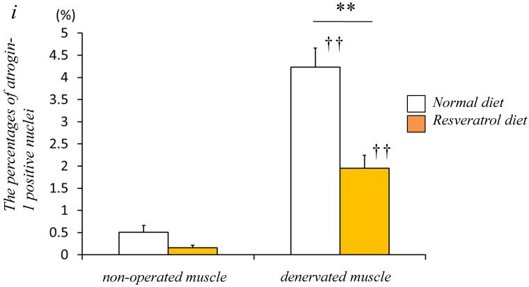
Figure 2.
Serial cryosections of the gastrocnemius muscle of mice fed normal and resveratrol diets. Atrogin-1 immunoreactivity was visualized using Rhodamine-conjugated antibody. In non-operated gastrocnemius muscle of both diets, immunofluorescence labeling showed that atrogin-1 was not present in the any nuclei of muscle fibers (a-d). Marked increases of atrogin-1 immunoreactivity were observed in several nuclei of denervated muscle of both mice (e-h). Densitometric analysis showed significant increasee in atrogin-1 immunoreactivity in the denervated muscle of mice fed between normal and resveratrol diets (i). However, the percentage of atrogin-1 positive nuclei were significantly lower in the mice fed the resveratrol diet than those of normal one (i). White circles and squares indicate the same fibers on different immunoimages. White arrows denote the atrogin-1-positive nuclei. Bar = 50 μm. ††: p < 0.01 compared with the non-operated muscle. **: p < 0.01 compared with normal diet.


