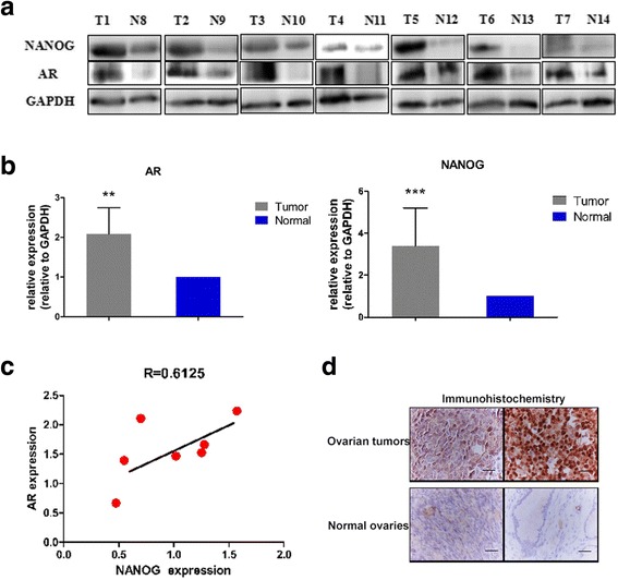
Fig. 1.

AR expression in ovarian tumors and corresponding Nanog gene expression. a) Western blot analysis of AR and Nanog expression in 7 ovarian tumors and 7 normal ovaries. b) AR and Nanog expression in ovarian tumors were higher than that in normal ovaries. Quality control software (Bio-Rad) was used to obtain the quantitative value relative to GAPDH. c) Correlation analysis of the AR and Nanog expression levels, indicating a correlation coefficient (R) of 0.61. d) Immunohistochemistry staining of AR expression in ovarian tumors and normal ovaries. In ovarian tumors, the positive (brown) staining is obvious. Bar value: 100 μM. T: ovarian tumor. N: normal ovaries. GAPDH: loading control. ** P < 0.01; *** P < 0.001
