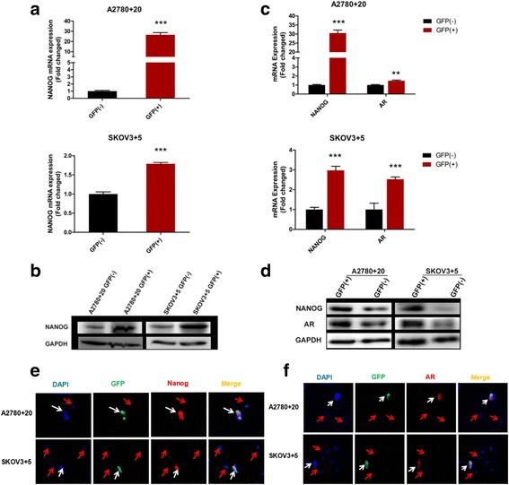
Fig. 3.

AR and Nanog co-exist in GFP Nanog (+) monoclonal cells. a and b) Nanog mRNA and protein expression in the Nanog GFP (+)/(−) cells of the A2780 and SKOV3 cell lines was examined by RT-qPCR and western blot, respectively. c and d) The mRNA and protein expression of both Nanog and AR in the Nanog GFP (+)/(−) cells of the A2780 and SKOV3 cell lines was also examined. e and f) Localization of Nanog and AR expression in Nanog GFP (+)/(−) cells determined by immunofluorescence staining; **P < 0.01; ***P < 0.001. GAPDH: loading control. Bar value: 100 μM. White arrow: double-positive cell. Red arrow: double-negative cell
