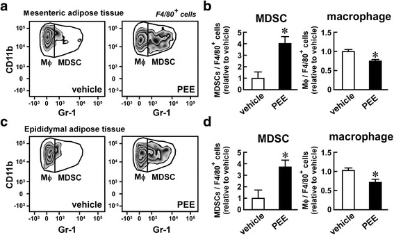
Fig. 2.

Effects of PEE on the number of macrophages and MDSCs in the visceral adipose tissue of high fat diet (HFD)-fed mice. HFD-fed C57BL/6 mice were intraperitoneally injected with PEE (100 mg/kg, twice weekly) or vehicle for one month. Subsequently, (a, b) mesenteric and (c, d) epididymal adipose tissue were subjected to FACS analysis of macrophages and MDSCs. a, c Representative FACS images of four replicates. Only F4/80+ living cells are shown. b, d Proportion of macrophages and MDSCs in F4/80+ myeloid cells from (b) mesenteric and (d) epididymal adipose tissue. Data represents means ± SD of four samples. *P < 0.05 vs. vehicle-treated mice
