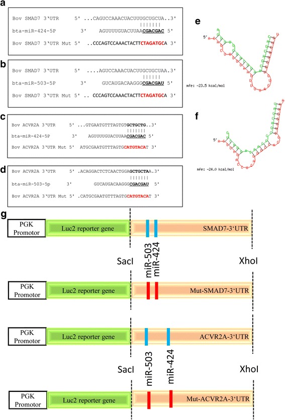
Fig. 1.

The miRNA-mRNA binding sites in bovine SMAD7 3´-UTR (a, b) and ACVR2A 3´-UTR sequences (c, d), Bold and underlined letters indicate putative binding sites and mutated regions. The minimum free energies (kcal/mol) of miR-424 (e) and miR-503 (f). Schematic diagram of the reporter constructs containing the putative miRNA-mRNA binding sites of the bovine SMAD7 and ACVR2A 3´-UTR sequences (g)
