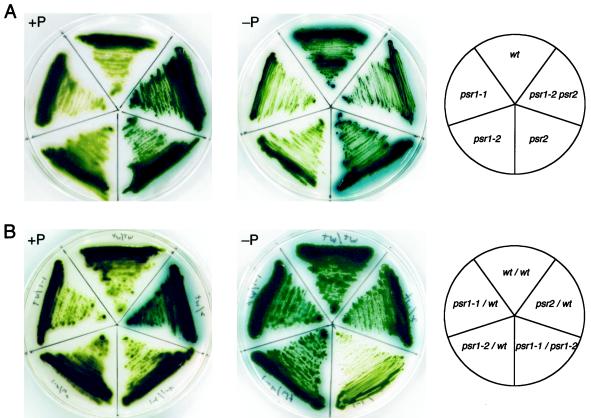
Figure 2.
Qualitative analysis of phosphatase activity secreted by wild-type cells, mutant strains, and vegetative diploids. Wild-type cells (wt), psr1-1, psr1-2, psr2, and psr1-2 psr2 (A) and vegetative diploids of wild-type and the various mutant strains (B) were streaked onto TAP (+P) and TA (−P) solid media before spraying the plates with the colorimetric phosphatase substrate X-Pi. The plates were allowed to develop for 2 h before recording the results. The template shows the positions of the various mutants (A, right) and the different vegetative diploids (B, right) on the plates.

