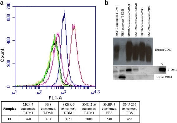
Fig. 2.

The T-DM1 and CD63 content of Type A exosomes. T-DM1-treated SKBR-3 and SNU-216 exosomes (red and blue, respectively) have a higher fluorescence intensity (FI) in flow cytometry indicating a higher T-DM1 content in these exosomes as compared with the control samples (T-DM1-treated MCF-7 exosomes, pink; T-DM1-treated FBS exosomes, green; PBS-treated SKBR-3 exosomes, orange; PBS-treated SNU-216 exosomes, black) (a). The human exosome marker protein CD63 is present in the Type A exosomes obtained from the culture media of the human cell lines, and the bovine CD63 exosome marker in FBS treated with T-DM1 in a Western blot analysis (b). T-DM1 content was high in SKBR-3 cell line-derived exosomes treated with T-DM1 (B). 55 ng of T-DM1 was used as a positive control (X)
