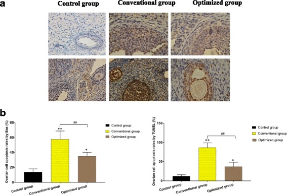
Fig. 3.

Apoptosis of ovarian cells in the different groups. a Representative ovarian images by Bax and TUNEL staining in the fresh group, conventional group and optimized group. (scale bar =50 μm). b The apoptosis rates of ovarian cells in different groups compared with the control group, *P < 0.05, **P < 0.01; compared with the conventional group, ##P < 0.01
