Fig. 1.

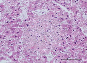
Photomicrograph showing liquefactive necrosis in the liver. Nuclear debris is seen as dark basophilic granules in the lesion. Yellow-necked mouse, HE. Bar = 100 µm

Photomicrograph showing liquefactive necrosis in the liver. Nuclear debris is seen as dark basophilic granules in the lesion. Yellow-necked mouse, HE. Bar = 100 µm