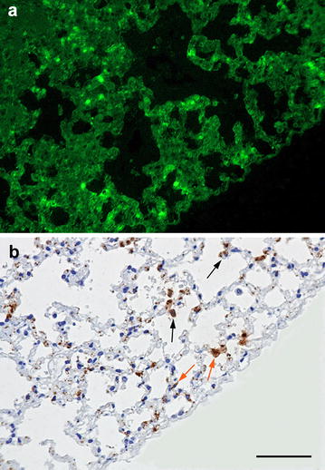
Fig. 2.

Microcopic visualization of F. tularensis in the lung of a yellow-necked mouse. a Indirect immunofluorescence photomicrograph of the lung infected by F. tularensis. F. tularensis bacteria are visualized in bright green fluorescence in the alveolar septa. b Immunohistochemistry for F. tularensis of the same area shown in a reveals their presence in the cytoplasm of pneumocytes (orange arrow) and alveolar macrophages (black arrow). Yellow-necked mouse. Bar = 100 µm
