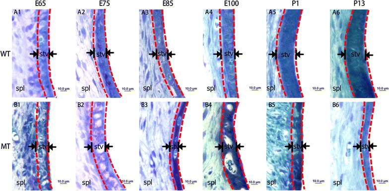
Fig. 1.

Malformation of the stria vascularis (SV) in developing cochleae of pigs with MITF-M mutation. A1–6 Illustrate the SV in the wild type cochleae from E65 to P13. B1–6 Illustrate the SVs in mutant pigs from E65 to P13. Images show that the SV of MITF-M mutant pigs are remarkably thinner than the normal pigs after E85. All images were taken from the basal turn of cochleae (Wild type, WT; mutant type, MT; spiral ligament, Spl; stria vascularis, stv. Bar = 10 μm)
