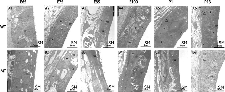
Fig. 2.

Lack of intermediate cells in the developing stria vascularis (SV) of MITF-M mutant pigs from E65 to P13. Since E75, the SV in the wild type cochleae was typically comprised of three layers: marginal, intermediate and basal cell layer (A1–6). In contrast, since E85 the SV in mutant type appeared to be extremely disorganized with absence of intermediate cells, and degeneration of marginal and basal cells (B1–6) (Wild type, WT; mutant type, MT; scala media, SM; marginal cells, pentacles; intermediate cells, asterisks; basal cells, triangles. Bar = 5 μm)
