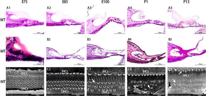
Fig. 3.

Defected organ of Corti and sensory hair cells in the cochleae of the MITF-M mutant pigs from E75 to P13 (B1–5). All images were taken from the basal turn of cochleae. The organ of Corti and sensory hair cells in the cochleae of WT pigs were manifested from E75,E85,E100,P1 and P13 (A1–5). The SEM image of hair cells in the cochleae of the MITF-M mutant pigs were manifested in C1–5. The degeneration of the organ of Corti in the MT cochlea was manifested from E100 and onwards (B3–5) as absence of inner and outer hair cells was observed (C3–5). All images were taken from the basal turn of cochleae. Wild type, WT; mutant type, MT; inner hair cells, IHCs; outer hair cells, OHCs. Bar in A1–B5 = 50 μm, Bar in C1–5 20 μm
