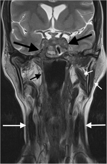
Fig. 3.

Coronal T2-weighted MRI image of the skull base and neck (pre-cyclophosphamide treatment). There is severe chronic inflammatory soft tissue abnormality affecting the cavernous sinuses and central and lateral skull base, including the left cavernous sinus and right sphenoid bone and foramen rotundum (large black arrows). Secondary cranial nerve palsies are present. Palsy of the mandibular division of the right trigeminal nerve has resulted in atrophy of the right masticator space muscle, in comparison to the more normal muscle volume on the left (short white arrows). Lower cranial nerve palsies are demonstrated by atrophy of the right superior pharyngeal constrictor muscle (short black arrow) and sternocleidomastoid muscles (long white arrows)
