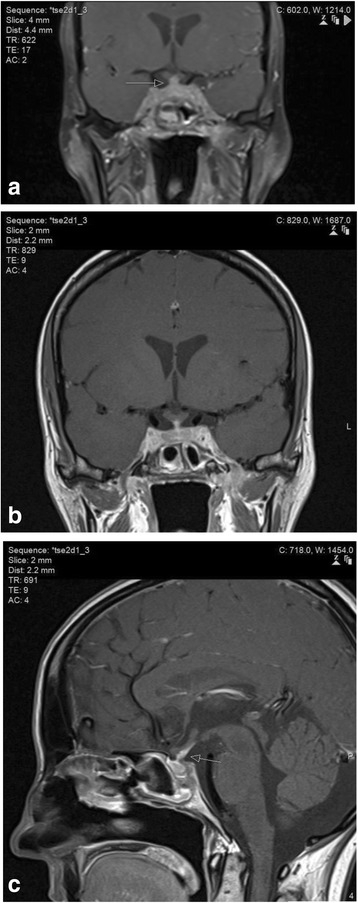
Fig. 4.

MR images of the brainstem, skull base and pituitary gland. (a) Coronal contrast-enhanced fat-saturated T1-weighted image of the central skull base demonstrates pathological enhancement of the cavernous sinus, pituitary gland and pituitary stalk (arrow). The abnormalities are non-discrete and radiologically inconsistent with a microadenoma or invasive macro adenoma of the pituitary gland. (b) Dedicated thin-section coronal contrast-enhanced T1-weighted image of the pituitary demonstrates the abnormal enhancement and thickening of the pituitary stalk as well as the cavernous sinus and pituitary parenchyma in more detail. (c) Dedicated thin-section sagittal contrast-enhanced T1-weighted image of the pituitary demonstrates the abnormal generalised thickening and enhancement of the pituitary stalk (arrow) consistent with granulomatous infundibulitis
