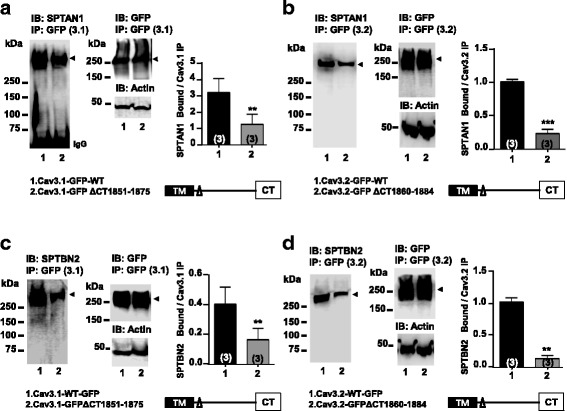
Fig. 2.

Effect of Cav3.1-GFP and Cav3.2-GFP C-terminal deletions on SPTAN1 and SPTBN2 binding in tsA-201 cells. (a) Cav3.1-GFP ∆CT (1851–1875) and wild type channel immunoprecipitates probed with anti-Spectrin αII (SPTAN1) polyclonal antibody. Densitometry analysis of SPTAN1 bound to Cav3.1-GFP immunoprecipitates is shown. (P = 0.0051, n = 3). (b) Cav3.2-GFP ∆CT (1860–1884) and wild type channel immunoprecipitates probed with anti-Spectrin αII (SPTAN1) polyclonal antibody. Densitometry analysis of SPTAN1 bound to Cav3.2-GFP immunoprecipitates is shown (P = 0.0005, n = 3). (c) Cav3.1-GFP ∆CT (1851–1875) and wild type channel immunoprecipitates probed with anti-Spectrin βII (SPTBN2) polyclonal antibody. Densitometry analysis of SPTBN2 bound to Cav3.1-GFP immunoprecipitates is shown (P = 0.0085, n = 3). (d) Cav3.2-GFP ∆CT (1860–1884) and wild type channel immunoprecipitates probed with anti-Spectrin βII (SPTBN2) polyclonal antibody. Densitometry analysis of SPTBN2 bound to Cav3.2-GFP immunoprecipitates is shown. (P = 0.0047, n = 3)
