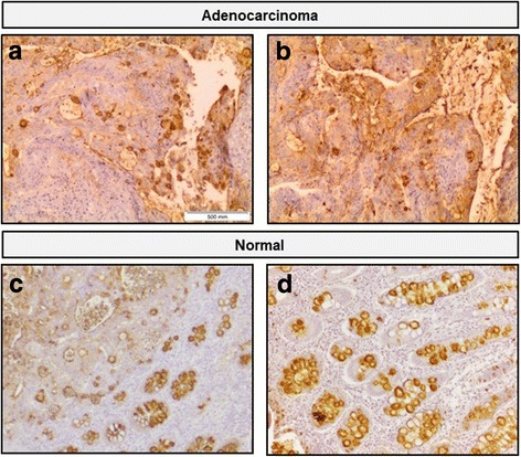
Fig. 4.

Immunohistochemistry staining of colon adenocarcinoma and normal colon tissue with E-Ig chimera and HECA-452 antibody. The E-Ig chimera that recognizes selectin ligands was used to stain colon adenocarcinoma (a) or normal colon (c) tissues, with 40× magnification. The HECA-452 antibody, that recognizes sLeX and sLeA glycans, was used to stain colon adenocarcinoma (b) or normal colon (d) tissues. In case of tumor tissue, images were also taken in sequences from the same tissue section of the same paraffin block of tumor tissue. Brown color indicates E-Ig or HECA-452 reactivity. In adenocarcinoma, the lamina propria showed E-Ig staining exclusively on nests of neoplastic cells, while HECA-452 staining showed positive scattered staining. In normal tissue, both E-Ig chimera and HECA-452 stains the lumens of the crypts and, in particular, the goblet cells
