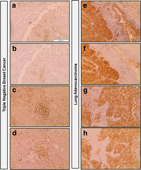
Fig. 5.

Immunohistochemistry staining of triple negative breast cancer and lung adenocarcinoma tissues with E-Ig chimera and HECA-452 monoclonal antibody. E-Ig was used for staining the E-selectin ligands in triple negative breast cancer (a and c) and in lung adenocarcinoma (e and g) tissues. sLeX and sLeA were stained with HECA-452 antibody in triple negative breast cancer (b and d) and in lung adenocarcinoma (f and h) tissues. Brown color indicates E-Ig or HECA-452 positive reactivity. Images were taken in sequences from the same tissue section of the same paraffin block of tumor tissue, with a 10× magnification
