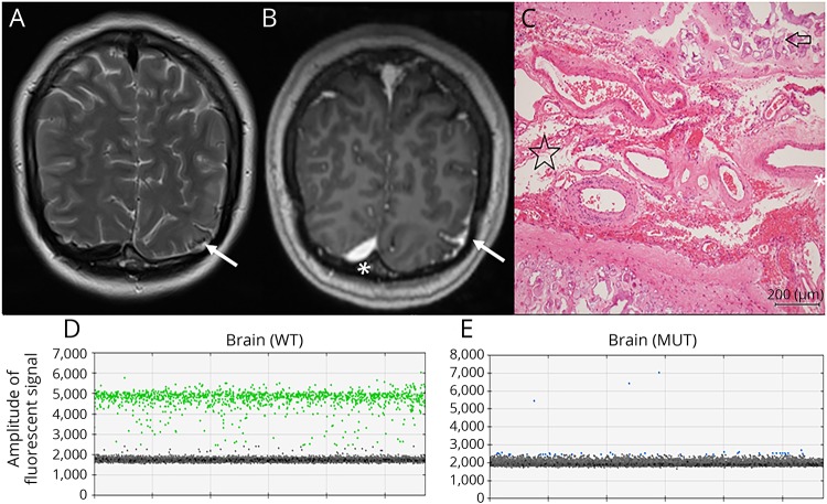
Figure 2. Imaging, histopathology, and molecular evaluation of case 3 with subtler MRI findings.
(A) Precontrast T2-weighted coronal MRI scan showing subtle signal change and calcification in the left occipital region (arrow) posteroinferiorly involving the occipital cortex or leptomeninges. Calcification was confirmed on CT (not shown). (B) Postcontrast T1-weighted coronal MRI scan showing leptomeningeal enhancement in the same region. Enhancement in the right occipital region (asterisk) is due to the normal transverse sinus. (C) Hematoxylin and eosin stained image showing subarachnoid angiomatosis (starred) between adjacent cerebral gyrae with cortical calcification (arrow). (D) Identification of the wild-type GNAQ allele (in green) in the brain by digital PCR. (E) Identification of the mutant GNAQ R183Q allele (in blue) in the brain. Droplets without genomic DNA templates are gray. Y-axis, amplitude of fluorescent signal. WT = wild-type GNAQ probe; MUT = mutant GNAQ R183Q probe.

