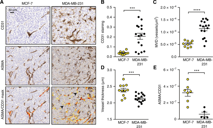
Fig. 3.
MDA-MB-231 exhibit a high-microvessel density but relatively poor maturity compared to MCF-7. a IHC representative micrographs for each tumour type stained with CD31 to mark endothelial cells and ASMA to mark the supporting pericyte layer (ASMA+ cells surrounding blood vessels). The lowest panel shows the mask used to count co-localised ASMA and CD31 staining on adjacent sections (orange overlap/yellow CD31+ only). Arrowheads indicate CD31+ with no ASMA staining in MDA-MB-231. Scale bar = 30 µm. MDA-MB-231 tumours show increased overall CD31 staining (b) and microvessel density (MVD, c), but decreased vessel thickness (d) and ASMA coverage (e) compared to MCF-7. For b, c and d, nMCF−7 = 12 and n231 = 16. For E, nMCF−7 = 7 and n231 = 4. All panels, data expressed as mean ± SEM. ***p < 0.001, ****p < 0.0001 by unpaired two-tailed t-test

