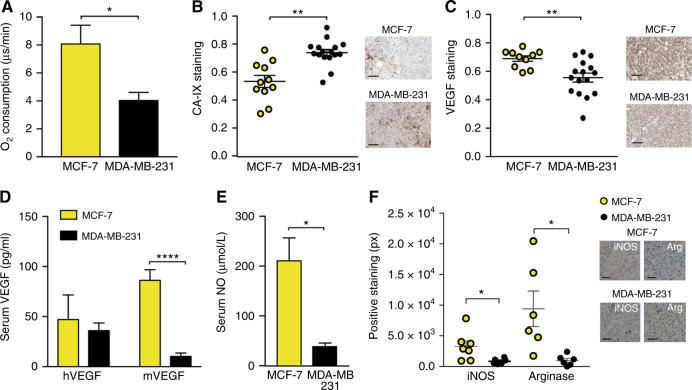
Fig. 4.
The hypoxic and inflammatory phenotype differs between the two breast tumour models. MDA-MB-231 cells have a lower oxygen consumption rate (a) yet show higher hypoxia in tumours (b). VEGF staining is lower in MDA-MB-231 tumours (c). Serum mouse VEGF (mVEGF, d) and nitric oxide (NO, e) are also lower in MDA-MB-231 tumours. f MCF-7 shows an inflammatory phenotype, with higher staining for iNOS and Arginase indicating the presence of type 1 and 2 macrophages respectively. Scale bars in (b) and (c) = 50 µm; f = 20 µm. For (b) and (c) nMCF−7 = 12 and n231 = 16. For (d) and (e) nMCF−7 = 6 and n231 = 9. All panels, data expressed as mean ± SEM, *p < 0.05, **p < 0.01, ***p < 0.001, ****p < 0.0001 by unpaired two-tailed t-test

